Career Overview
Explore My Work
Since the beginning of my journey in this field, my dedication to research and development and my motivation to learn and innovate have led me to work on exceptional projects and with enthusiastic and beautiful minds.
I am grateful for a profession that I’m truly passionate about, and I am proud to share samples of some of my greatest achievements.
Take a moment to check out my portfolio, and feel free to get in touch if you have any questions.
Passion: Computer Graphics, Computer Vision, Augmented / Virtual / Mixed Reality, Artificial Intelligence, Data Visualisation, Data Science, Medical Imaging, Human-Computer Interactions, Internet of Things, Psychology, Philosophy, Neuroscience, Physics, Engineering
"Tomorrow is less to discover than to invent"
— Gaston Berger


Publications
List of publications in peer reviewed journals and conference proceedings
Creating 3D worlds through storytelling and narration
While contemporary game engine tools have allowed a lower barrier of entry into creating 3D environments, many rely on engineering, architectural or cartographic approaches. While useful, this usually facilitates very particular methods of 3D space creation. In this paper, we present our tool for creating 3D spaces through the use of narration and storytelling as an interface. Our platform utilizes voice recognition to allow users to narrate a story or event and have elements of the story appear in 3D space as virtual objects in real-time. Through further voice commands or the use of drag-and-drop controls, users can then reconfigure these objects to better suit their story or create new variations of the story. Our platform is designed for immersive systems such as VR HMDs, 3D-enabled cylindrical screens, stereo walls, and non-immersive ones such as desktops and tablets allowing for accessibility and collaboration. A novel contribution of this paper is this method of building worlds through narration. To provide some insight into its capabilities and the reasoning behind design decisions, an account of constructing the story Lessons in Vinyl within the tool during early development is presented.
Authors: Cameron Edmond, Dominic Branchaud, Tomasz Bednarz
Publication: OzCHI '20: 32nd Australian Conference on Human-Computer-Interaction, December 2020
Digital Twin of the Australian Square Kilometre Array (ASKAP)
In this work, we present the Digital Twin of the Australian Square Kilometre Array Pathfinder (ASKAP) - an extended reality framework for telescope monitoring. Currently, most of the immersive visualisation tools developed in astronomy primarily focus on educational aspects of astronomical data or concepts. We extend this paradigm, allowing complex operational network controls with the aim of combining telescope monitoring, processing and observational data into the same framework.
Authors: Tomasz Bednarz, Dominic Branchaud, Florence Wang, Justin Baker, Malte Marquarding
Publication: Siggraph Asia '20, December 2020, Article No.: 15, Pages 1
Double Vision: Digital Twin applications within Extended Reality
The purpose of this project was to create a Digital Twin (DT) of the Expanded Perception and Interaction Centre (EPICentre), using Extended Reality to better visualise Internet of Things (IoT) sensor data such as temperature and humidity throughout the building. An immersive application was developed for multiple platforms, allowing users to interact with the DT model through Windows Mixed Reality Platforms, Oculus Platforms and a Cylindrical Screen. This project lends itself as an engaging introduction into DTs through the immersive experience of data analytics, generalised for the public using familiar sensors-driven datasets.
Authors: Jade Jiang, Michael Tobia, Robert Lawther, Dominic Branchaud, Tomasz Bednarz
Publication: SIGGRAPH '20 Appy Hour: Special Interest Group on Computer Graphics and Interactive Techniques Conference Appy Hour, Virtual Event, USA, August 2020
Immersive Analytics using Augmented Reality for Computational Fluid Dynamics Simulations
This project aimed to employ multi-sensory visual analytics to a Computational Fluid Dynamics (CFD) dataset using augmented and mixed reality interfaces. Initial application was developed for a Hololens which allows users to interact with the CFD data using gestures, enabling control over the position, rotation and scale of the data, sampling, as well as voice commands that provide a range of functionalities such as changing a parameter or render a different view. This project leads to a more engaging and immersive experience of data analysis, generalised for CFD simulations. The application is also able to explore CFD datasets in fully collaborative ways, allowing engineers, scientists, and end-users to understand the underlying physics and behaviour of fluid flows together.
Authors: Tomasz Bednarz, Michael Tobia, Huyen Nguyen, Dominic Branchaud
Publication: VRCAI '19: The 17th International Conference on Virtual-Reality Continuum and its Applications in Industry November 2019 Article No.: 46 Pages 1–2
Massive Networks - Visualizing Very Large-Scale Graphs in Immersive Environments
This work presents our strategy and pipeline architecture for visualizing very large-scale graphs in an immersive environment, using a high-performance graphics approach. The innovation lies in utilizing GPUs for real-time cluster-based interactive rendering, but also intermediate graph representation that utilizes Khronos Group's GLTF file format, and interaction design.
Authors: Daniel Filonik, Dominic Branchaud, Robert Lawther, Piotr Szul, Alex Collins, Tomasz Bednarz
Publication: HPG'18, August 2018, Vancouver, Canada
Visual microscope for massive genomics datasets, expanded perception and interaction
An innovative fully interactive and ultra-high resolution navigation tool has been developed to browse and analyse gene expression levels from human cancer cells, acting as a visual microscope on data. The tool uses high-performance visualisation and computer graphics technology to enable genome scientists to observe the evolution of regulatory elements across time and gain valuable insights from their dataset as never before.
Authors: Dominic Branchaud, Walter Muskovic, Maria Kavallaris, Daniel Filonik, Tomasz P Bednarz
Publication: SIGGRAPH '18: ACM SIGGRAPH 2018 Posters August 2018 Article No.: 73 Pages 1–2
3D shape reconstruction of bone from two x-ray images using 2D/3D non-rigid registration based on moving least-squares deformation.
Several studies based on biplanar radiography technologies are foreseen as great systems for 3D-reconstruction applications for medical diagnoses. This paper proposes a non-rigid registration method to estimate a 3D personalized shape of bone models from two planar x-ray images using an as-rigid-as-possible deformation approach based on a moving least-squares optimization method. Based on interactive deformation methods, the proposed technique has the ability to let a user improve readily and with simplicity a 3D reconstruction which is an important step in clinical applications. Experimental evaluations of six anatomical femur specimens demonstrate good performances of the proposed approach in terms of accuracy and robustness when compared to CT-scan.
Author(s): T. Cresson; D. Branchaud; R. Chav; B. Godbout; J. A. de Guise
Publication: Proc. SPIE 7623, Medical Imaging 2010: Image Processing, 76230F (12 March 2010);
Coupling 2D/3D registration method and statistical model to perform 3D reconstruction from partial x-rays images data.
3D reconstructions of the spine from a frontal and sagittal radiographs is extremely challenging. The overlying features of soft tissues and air cavities interfere with image processing. It is also difficult to obtain information that is accurate enough to reconstruct complete 3D models. To overcome these problems, the proposed method efficiently combines the partial information contained in two images from a patient with a statistical 3D spine model generated from a database of scoliotic patients. The algorithm operates through two simultaneous iterating processes. The first one generates a personalized vertebra model using a 2D/3D registration process with bone boundaries extracted from radiographs, while the other one infers the position and the shape of other vertebrae from the current estimation of the registration process using a statistical 3D model. Experimental evaluations have shown good performances of the proposed approach in terms of accuracy and robustness when compared to CT-scan.
Authors: Cresson T, Chav R, Branchaud D, Humbert L, Godbout B, Aubert B, Skalli W, De Guise JA.
Publication: Conf Proc IEEE Eng Med Biol Soc. 2009; 2009:1008-11.
Fast 3D reconstruction of the spine from biplanar radiography: a diagnosis tool for routine scoliosis diagnosis and research in biomechanics
Reconstruction methods from biplanar radiography allow a 3D clinical analysis, for patients in standing position, with a low radiation dose. This low-dose and postural imaging modality is thus very interesting for scoliosis clinical diagnosis and research in biomechanics. Nevertheless, such applications require both accurate and fast 3D reconstruction methods.
Fast approaches, based on statistical models (Pomero et al. 2004; Gille et al. 2007), allow to obtain an estimate of the spinal 3D reconstruction within 14 min. However, this remains too tedious to be used in a clinical routine. The purpose of this study is to propose and evaluate a novel semi-automated 3D reconstruction method of the spine from biplanar radiography. This method relies on a parametric model of the spine using statistical inferences and automatic registration methods based on image processing. Two reconstruction levels are proposed: a first reconstruction level (‘Fast Spine’), providing a fast estimate of the 3D reconstruction and accurate clinical measurements, dedicated to a routine clinical use, and a more accurate second reconstruction level (‘Full Spine’) for applications in biomechanical research.
Authors: L. Humbert, J.A. De Guise, B. Godbout, T. Cresson, B. Aubert, D. Branchaud, R. Chav, P. Gravel, S. Parent, J. Dubousset, W. Skalli
Publication: Computer Methods in Biomechanics and Biomedical Engineering; Vol. 12, No. S1, August 2009, 151-152
Performing fast 3D reconstruction of the knee from biplanar x-ray images through moving least squares deformation for surgical planning.
Modeling anatomical structures and calculation of 3D measurements such as angles and distances are becoming crucial therapeutical tools in computer assisted surgery systems for clinical evaluation. Nowadays, the main goal is to generate the patient-specific 3D bone reconstructions and measurements avoiding exposition to high doses of radiations and expensive treatments. Several studies based on biplanar radiography technologies are foreseen as an alternative to Magnetic Resonance Imaging (MRI) and Computerized Tomography (CT) in orthopaedic applications for many standard diagnoses where the standing position is essential. 3D modeling methods, using only few x-ray images, are mainly based on image processing techniques with a template model using the statistical shape analysis of a collection of related anatomical structures. However, these methods require a lot of data describing the considered structure. Other approaches use an elastic deformation applied to a template model without considering any statistical information. The kriging algorithm is used to fit the template model on contours previously extracted from images.
We use the moving least squares (MLS) deformation technique to achieve fast detail-preserving deformation, for anatomical surface reconstruction from two radiographs obtained in a calibrated system. This study presents an interactive system based on the MLS method to rapidly evaluate the shape and the 3D alignment of the lower limb using biplanar x-rays images with the patient in the load-bearing standing position.
Authors: Cresson, T., Godbout, B., Branchaud, D., Chav, R., Chaibi, Y., Aubert, B., Skalli, W. et de Guise, JA.
Publication: International Journal of Computer Assisted Radiology and Surgery, vol. 4, nº suppl. 1. p. 113-114
Surface reconstruction from planar x-ray images using moving least squares.
Planar radiographs still are the gold standard for the measurement of the skeletal weight-bearing shape and posture. In this paper, we propose to use an as-rigid-as-possible deformation approach based on moving least squares to obtain 3D personalized bone models from planar x-ray images. Our prototype implementation is capable of performing interactive rate shape editing. The biplane reconstructions of both femur and vertebrae show a good accuracy when compared to CT-scan.
Authors: T. Cresson; B. Godbout; D. Branchaud; R. Chav; P. Gravel; J.A. De Guise
Publication: 2008 30th Annual International Conference of the IEEE Engineering in Medicine and Biology Society, Vancouver, BC, 2008, pp. 3967-3970
Spinal Vertebrae Edge Detection by Anisotropic Filtering and a local Canny-Deriche Edge Detector
This paper describes a robust method for spinal vertebrae edge detection from biplanar X-ray radiographic images (lateral and antero-posterior). It uses anisotropic filtering and a Canny-Deriche edge detector applied locally and in a region of interest. The anatomical structure of interest in this work is the spinal area. Anisotropic filtering is applied so as to preserve edge structures while reducing image noise. The proposed technique has been validated on a database of X-ray radiographic images obtained from the EOS system. Results are compared to the edges drawn by a medical expert.
Authors: N. Mezghani, S. Deschˆenes, B. Godbout, D. Branchaud and J.A. de Guise
"Do not go where the path may lead. Instead, go where there is no path and leave a trail"
— Ralph Waldo Emerson


Patents
High impact quality creative work
List of notable patented innovations
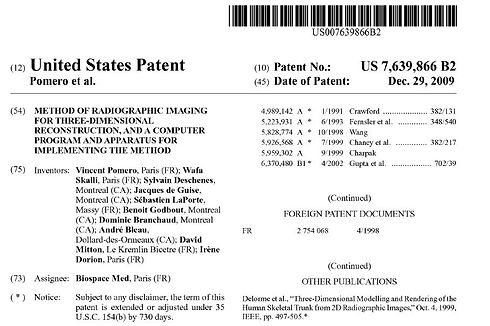
Method of radiographic imaging for three-dimensional reconstruction, and a computer program and apparatus for implementing the method
Patent number: US7639866B2
A radiographic imaging method for three-dimensional reconstruction in which the three-dimensional shape of a model representing an object is calculated from a geometrical model of the object that is known a priori, and obtained from a confinement volume of the object estimated from a geometrical pattern visible in two images and from knowledge of the positions of the sources. A geometrical model is used that comprises information making it possible using an estimator for the object to establish a geometrical characteristic for the model representing the object.
Cited by: General Electric Co, Philips Electronics, Toshiba Corp, Fuji Photo Film Co Ltd, Siemens Corp Research Inc., Hitachi Ltd, University of North Carolina, University of California, University of Waseda, Research Foundation the State Univ. of New York, etc.
Alt Link: https://patents.google.com/patent/US7639866

Radiographic imaging method for three-dimensional reconstruction, device and computer program for implementing said method
Patent number: FR2856170B1
The inventive radiographic imaging method for three-dimensional reconstruction consists in computing a three-dimensional form of an object representing model which is based on the already known geometrical form of said object obtainable from the confining volume thereof by evaluating a geometric pattern visible on two pictures and a source position. The geometric model containing data which makes it possible to set up a geometric characteristic of the object representing model by means of the object estimator is used.
Cited by: Tokyo Shibaura Electric Co, Nippon Telegr & Teleph Corp (NTT), Thomson Consumer Electronics Inc., Matsushita Electric Ind Co Ltd, Meidensha Corp, Univ. Joseph Fourier, Biospace Instr, Hologic Inc., Orthosoft Inc., BrainLAB AG, Rapiscan Systems Inc., etc.

Patent number: US20100241405A1
The present invention is related to a method for reconstruction of a three-dimensional model of an osteo-articular structure, e.g. human spine, based on two-dimensional patient-specific detection data of the structure, e.g. two calibrated radiographs, and on a previously established preliminary solution corresponding to a solution model of the structure. The preliminary solution initially comprises a priori knowledge of the structure, established from structures of the same type. The preliminary solution is further modified using the two-dimensional patient-specific detection data.
Cited by: Univ Joseph Fourier, Framatome SAS, École Nationale Supérieure des Arts et Métiers, Conseil National de la Recherche Scientifique, Brainlab AG, Biospace Instr, EOS Imaging, William Marsh Rice University, Université Laval, etc.
Alt link: https://app.dimensions.ai/details/patent/US-9142020-B2


